

Discover why serum creatine kinase variability makes it an imperfect single marker for muscle microtrauma. We compare blood biomarkers with functional power output recovery tests.
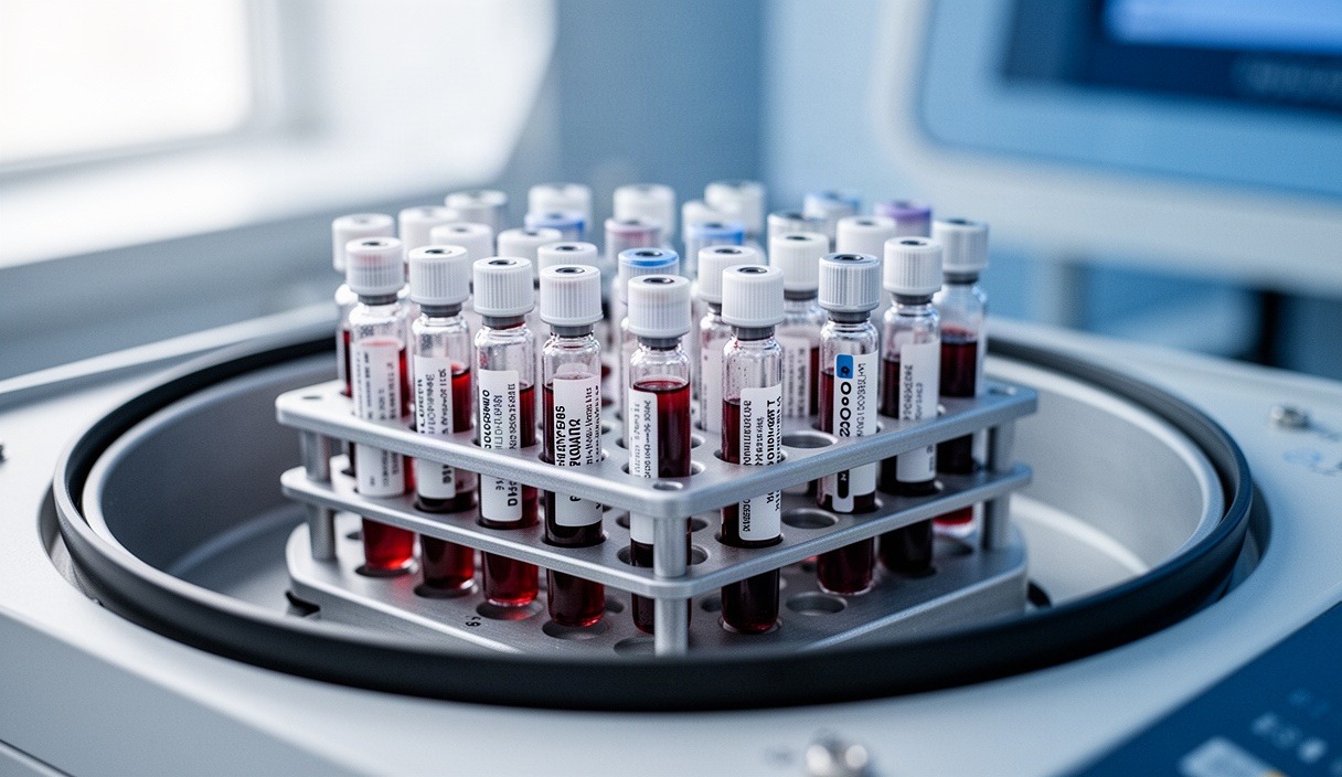
Athletic performance staff and strength practitioners frequently order venous blood panels or point-of-care capillary tests to quantify exercise-induced muscle damage. Among the measurable circulating proteins, serum creatine kinase (CK) remains the most common metric. Released into the bloodstream after structural microtrauma, creatine kinase serves as a direct indicator of cellular membrane disruption. However, its translation from a laboratory value into an actionable training adjustment is rarely straightforward.
Relying on creatine kinase as an absolute indicator of recovery creates serious blind spots in a performance program. Serum concentrations often diverge significantly from an athlete's physical work capacity, local soreness, and histological recovery status. Understanding the biological mechanisms of membrane leakage, clearance pathways, and the massive inter-individual variability between athletes is necessary to determine whether a spike in blood values warrants modifying a training load or represents a normal biological response to mechanical tension.
Serum Creatine Kinase Leakage Mechanisms
Creatine kinase is an intracellular enzyme found predominantly in skeletal muscle, cardiac muscle, and the brain. In skeletal tissue, it catalyzes the reversible transfer of a high-energy phosphate group between adenosine triphosphate (ATP) and phosphocreatine. Under normal resting conditions, the intact sarcolemma (the muscle cell membrane) acts as a secure barrier, maintaining cytosolic proteins inside the fiber and keeping baseline serum levels within a relatively modest range, typically between 40 and 250 U/L for non-exercising individuals.
Mechanical stress disrupts this containment. High-force eccentric actions force active muscle fibers to lengthen while generating tension. This process shears structural proteins, particularly titin and desmin, and causes focal tears in the lipid bilayer of the sarcolemma. The resulting microtrauma disrupts intracellular calcium balance. Excess extracellular calcium floods the cytosol, activating calpains, which are calcium-dependent neutral proteases that digest structural proteins from within. This secondary enzymatic degradation increases sarcolemmal permeability.
Once the cell membrane is compromised, creatine kinase escapes from the sarcoplasm into the interstitial fluid. From the interstitium, the enzyme is taken up by the lymphatic drainage system rather than diffusing directly into local capillaries. This lymphatic transport path explains why blood levels do not peak immediately following a training bout. Creatine kinase enters systemic circulation through the thoracic duct, causing circulating concentrations to rise gradually. The peak typically appears between 24 and 72 hours post-exercise, though unaccustomed downhill running or maximal eccentric lifting can push this peak past 96 hours.
Clearance from systemic circulation is governed largely by the reticuloendothelial system, primarily via macrophages in the liver and spleen. Because the half-life of circulating creatine kinase ranges from 24 to 36 hours, clearance lag can keep blood values elevated long after the primary mechanical damage has stopped. The rate of this process depends on liver perfusion, the athlete's hydration status, and their systemic clearance capacity.
Inter-Individual Variability and High-Responder Phenomenon
The greatest challenge in interpreting creatine kinase data is the absence of a universally applicable reference range for athletes. Two teammates performing an identical lower-body eccentric protocol can produce vastly different blood concentrations. One may record an unremarkable rise to 420 U/L, while the other exceeds 14,000 U/L without displaying signs of functional impairment or abnormal distress. This disparity has led researchers to classify subjects into low, normal, and high responders.
Several physiological and structural factors drive this divergence:
- Genetic Polymorphisms: Variations in genes such as ACTN3 (which encodes alpha-actinin-3 in fast-twitch fibers) and MLCK (myosin light chain kinase) alter sarcomeric stability. Athletes homozygous for the ACTN3 577X null allele often display higher baseline membrane fragility and amplified enzymatic leakage following novel loading.
- Sex Differences: Circulating estrogen exerts a protective, membrane-stabilizing effect on lipid bilayers. As a consequence, female athletes frequently show lower absolute creatine kinase spikes than males exposed to identical relative workloads, although their subjective recovery timelines and structural damage can be identical.
- Muscle Mass and Fiber Architecture: Athletes with greater total skeletal muscle mass possess a larger reservoir of intracellular enzymes. Fast-twitch type II fibers are far more susceptible to eccentric damage than type I fibers. Athletes with a high proportion of fast-twitch motor units routinely produce higher enzyme readings.
- Prior Exposure and the Repeated Bout Effect: A single exposure to eccentric loading initiates structural adaptations, including sarcolemmal reinforcement and sarcomere genesis in series. This repeated bout effect can suppress enzyme leakage on subsequent sessions by more than 70 percent compared to the initial stimulus.
| Classification | Typical Post-Damage Peak Range | Time to Peak Clearance | Primary Physiological Drivers |
|---|---|---|---|
| Low Responder | 300 - 950 U/L | 24 to 48 hours | High slow-twitch distribution, established training history, higher relative estrogen. |
| Normal Responder | 1,000 - 4,500 U/L | 48 to 72 hours | Mixed fiber typing, moderate training age, standard sarcolemmal membrane stability. |
| High Responder | 5,000 - 28,000+ U/L | 72 to 120+ hours | Fast-twitch dominance, ACTN3 null variants, novel eccentric volume, large muscle volume. |
Because of these discrepancies, comparing raw creatine kinase numbers between athletes is practically useless. An isolated reading of 6,200 U/L in an elite sprinter might represent an ordinary post-session adaptation, whereas the same value in an endurance runner could point to significant, unmanageable tissue breakdown. Reliable interpretation requires comparing an individual athlete against their own historical baseline across identical training blocks.
Myoglobin and Lactate Dehydrogenase as Secondary Markers
To offset the limitations of using creatine kinase in isolation, sports scientists often track secondary blood biomarkers to construct a clearer profile of tissue breakdown. The two most common secondary markers are myoglobin and lactate dehydrogenase (LDH).
Myoglobin is a single-chain globular protein responsible for storing oxygen within myocytes. It has a molecular mass of roughly 17 kDa, making it significantly smaller than the 82 kDa creatine kinase dimer. Because of this smaller profile, myoglobin diffuses out of damaged sarcolemmal pores and interstitial spaces far more rapidly. Circulating myoglobin levels frequently spike within 1 to 4 hours post-exercise, reaching peak concentrations between 8 and 14 hours. It also clears quickly through glomerular filtration in the kidneys, returning to baseline within 24 to 36 hours if hydration is adequate.
The speed of myoglobin kinetics makes it a sensitive marker for acute, same-day trauma, whereas creatine kinase reflects lingering structural permeability. However, excessive myoglobin concentrations place a heavy burden on renal function. Under acidic physiological conditions, myoglobin precipitates in the renal tubules, which can lead to cast formation and acute tubular necrosis. When blood tests show extreme myoglobin elevations combined with concentrated, dark urine, the issue has moved beyond standard training soreness and requires immediate evaluation by a physician.
Lactate dehydrogenase acts on the opposite end of the temporal spectrum. This tetrameric enzyme is involved in cellular glycolysis, converting pyruvate to lactate. Muscle tissue primarily contains the LDH-4 and LDH-5 isoenzymes. Because of its large size (approximately 140 kDa), LDH takes longer to clear the lymphatic barrier. Serum LDH levels typically rise 24 to 48 hours following severe loading and remain elevated for up to 6 or 7 days. While non-specific on its own, an elevation in LDH alongside creatine kinase confirms that the initial inflammatory response remains active in the systemic phase.
| Biomarker | Molecular Mass | Earliest Detectable Rise | Peak Elevation Window | Return to Baseline |
|---|---|---|---|---|
| Myoglobin | 17 kDa | 1 to 2 hours | 8 to 14 hours | 24 to 36 hours |
| Creatine Kinase | 82 kDa | 6 to 12 hours | 24 to 72 hours | 3 to 6 days |
| Lactate Dehydrogenase | 140 kDa | 12 to 24 hours | 48 to 96 hours | 6 to 8 days |
Objective Muscle Function Tests Versus Blood Biomarkers
The strongest argument against relying solely on blood work is the frequent dissociation between enzyme leakage and an athlete's physical capacity. Muscle damage fundamentally impairs three performance characteristics: maximal force generation, rate of force development (RFD), and stretch-shortening cycle (SSC) efficiency. Multiple studies demonstrate that circulating enzyme levels correlate poorly with the magnitude of acute force loss and the subsequent speed of neuromuscular recovery.
For example, an athlete who performs a high-volume eccentric training session might experience an immediate 32 percent drop in maximal voluntary isometric contraction (MVIC) force. This decline occurs because disrupted cross-bridge cycles and excitation-contraction coupling failures, specifically calcium release failure from the sarcoplasmic reticulum, prevent the muscle from generating mechanical output. By hour 48, the athlete's MVIC may have recovered to 93 percent of baseline, even as their serum creatine kinase values hit a dramatic peak of 7,800 U/L due to delayed clearance kinetics.
Conversely, subjective ratings of delayed-onset muscle soreness (DOMS) dissociate from both blood markers and functional force output. Nociceptive pain peaks in response to inflammation within the epimysium, perimysium, and extracellular matrix, rather than the disruption of individual myofibrils. An athlete may feel debilitating soreness with only minor enzyme leakage, or report zero local tenderness while carrying significant sarcolemmal microtrauma.
Because of this, neuromuscular and functional tests serve as the ultimate arbiters of exercise readiness:
- Maximal Voluntary Isometric Contraction: Isometric mid-thigh pulls or isometric squats measure pure contractile force output without the eccentric-induced fatigue of repeated jumping. A deficit greater than 10 percent from baseline indicates unresolved excitation-contraction coupling failure.
- Countermovement Jump (CMJ) Kinetics: While jump height alone can be masked through motor control compensations, force plate metrics tell a clearer story. A prolonged eccentric phase duration, reduced eccentric braking RFD, and a depressed dynamic strength index directly highlight residual structural fatigue.
- Drop Jump and Reactive Strength Index (RSI): The fast stretch-shortening cycle (ground contact time under 250 milliseconds) is sensitive to reductions in muscle-tendon stiffness. Reductions in RSI highlight microtrauma in the series elastic elements that circulating blood panels cannot quantify.
A Practical Checklist for Determining Session Readiness
To avoid misinterpreting isolated blood markers, performance teams should use an integrated screening process. Work through this checklist prior to programming high-intensity or heavy-volume training:
- Compare Blood Metrics to Individual Baselines: If venous or point-of-care capillary values are available, compare current creatine kinase and myoglobin numbers against that specific athlete's historical mean under comparable training loads. Look for relative deviations rather than comparing the data against clinical laboratory reference ranges.
- Conduct a Hydration and Urine Color Screening: Ensure blood values are not artificially concentrated by hypohydration. Use a refractometer to verify that urine specific gravity is under 1.020. If dark or brown-tinted urine occurs alongside an enzyme surge, immediately suspend training and consult a medical professional to protect renal health.
- Evaluate Joint Range of Motion and Passive Stiffness: Measure passive and active joint range using a goniometer or an established mobility screen. Significant reductions in terminal joint angles (such as knee flexion following heavy eccentric hamstring work) indicate intracellular edema and protective tone.
- Assess Central and Autonomic Nervous System State: Measure resting heart rate variability (HRV) upon waking. A significant reduction in the root mean square of successive differences (RMSSD), or an atypical increase in resting heart rate exceeding 7 beats per minute, indicates systemic sympathetic dominance and incomplete recovery.
- Perform a Standardized Mechanical Force Test: Collect baseline kinetic data using an isometric pull or dynamic countermovement jump on calibrated force plates. If eccentric braking RFD or jump height remains depressed by more than 8 percent, mechanical recovery is incomplete, regardless of low blood enzyme numbers.
- Execute Progressive Specific Warm-Up Sets: Track bar speed or velocity-based training metrics during initial warm-up sets. If mean concentric velocity drops more than 0.08 meters per second at standard submaximal loads (such as 60 percent of 1RM), down-scale total working volume for the day.
Common Mistakes in Monitoring Muscle Damage
Coaches and sports science practitioners regularly make predictable errors when adding biomarker tracking to their workflow. The most frequent errors include:
- Using Creatine Kinase as a Clearance Marker for Competition: High serum levels often persist long after contractile capacity and power output have returned to baseline. Scratching an athlete based solely on an elevated number can lead to benching a fully functional performer.
- Failing to Account for Muscle Group Size: A maximal eccentric session targeting small muscle groups (such as the biceps brachii) induces acute local tenderness and functional drop-offs, but produces only a minor serum enzyme increase. Conversely, full-body eccentric work generates massive systemic leakage even if individual muscles recover quickly.
- Drawing Blood Samples at Inconsistent Times: Drawing blood immediately post-workout captures metabolic stress, not structural membrane damage. Conversely, testing at 24 hours one week and 72 hours the next renders the data incomparable because of the shifting curve of enzyme efflux.
- Ignoring Cross-System Medical Symptoms: Athletes with early-stage rhabdomyolysis or severe unaccustomed systemic breakdown often display normal resting vitals during the initial 12 hours. Never assume a high responder is fine simply because they feel capable of lifting. Always review hydration, systemic temperature, and renal indicators.
Next Steps for Implementation
To integrate muscle damage assessments into a training program, begin by developing consistent baseline values. During a low-stress preparatory week, collect two to three resting morning blood samples from each athlete to establish their unique personal reference range for creatine kinase and, if feasible, myoglobin.
Next, pair biomarker testing with a practical, objective functional measurement. Deploy dual force plates or a linear position transducer to record jumping mechanics or bar speed twice weekly. When a reading deviates by more than two standard deviations from the athlete's baseline, adjust the upcoming session by swapping high-velocity eccentric exercises for concentric-focused work, isometric training, or active recovery.
Finally, establish clear safety protocols with your medical staff. Define strict thresholds for clinical intervention, such as dark-colored urine, local swelling accompanied by numbness, or creatine kinase levels climbing past standard high-responder thresholds. While blood testing provides valuable insight into structural stress, it should always be interpreted alongside objective force production and skilled clinical oversight.
This publication provides educational information and does not constitute medical advice; consult a sports physician or licensed physical therapist for clinical guidance. Disclaimer